Archivo:Paramecium.jpg
De Wikipedia, la enciclopedia encyclopedia
Ver la imagen en su resolución original (751 × 738 píxeles; tamaño de archivo: 186 kB; tipo MIME: image/jpeg)
|
|
Este es un archivo de Wikimedia Commons, un depósito de contenido libre hospedado por la Fundación Wikimedia. Más abajo se reproduce su página de descripción con la información sobre su origen y licencia. |
Resumen
| DescripciónParamecium.jpg |
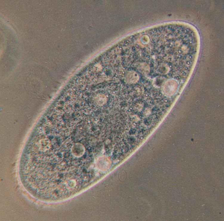
Deutsch: Paramecium aurelia - Optisches Mikroskop. Paramecium aurelia, der bekannteste von allen ciliaten. Die Blasen innerhalb der Zelle sind Vakuolen. Die gesamte Oberfläche ist mit Wimpern umgeben, die durch ihre schnelle Bewegung verwischt werden.
English: Paramecium aurelia. Optical microscope. Paramecium aurelia, the best known of all ciliates. The bubbles throughout the cell are vacuoles. The entire surface is covered in cilia, which are blurred by their rapid movement.
Français : Paramecium aurelia. Microscope optique. Le plus connu des ciliés. Les bulles que vous voyez sont des vacuoles. Tout le corps est couvert par des cils, qui sont flous sur l'image à cause de leurs mouvements rapides.
Polski: Paramecium aurelia - pantofelek, najbardziej znany ze wszystkich orzęsków. Bąbelki w środku komórki to wodniczki. Cała powierzchnia pantofelka pokryta jest rzęskami, które są na fotografii zamazane ze względu na ich szybki ruch.
Српски / srpski: Paramecium aurelia, najpoznatiji od svih trepljara pod optičkim mikroskopom. "Mehurići" u ćeliji paramecijuma su vakuole. Cela površina tela je prekrivena trepljama, koje su na slici mutne zbog toga što se brzo pokreću.
Türkçe: Paramecium aurelia - optik mikroskop. Paramecium aurelia, tüm siliyalılar içinde en çok bilinen türdür. Hücre boyunca yuvarlak olarak izlenen oluşumlar, vakuollerdir. Hücrenin tüm yüzeyi, hızlı hareketlerinden dolayı bulanık görüntü vermiş olan siliya ile kaplıdır. |
| Fecha | |
| Fuente | Originally uploaded to the English Wikipedia, where it was made by Barfooz. |
| Autor | Barfooz at the English Wikipedia. |
| Otras versiones | Transparent |
Licencia

|
Se autoriza la copia, distribución y modificación de este documento bajo los términos de la licencia de documentación libre GNU, versión 1.2 o cualquier otra que posteriormente publique la Fundación para el Software Libre; sin secciones invariables, textos de portada, ni textos de contraportada. Se incluye una copia de la dicha licencia en la sección titulada Licencia de Documentación Libre GNU.http://www.gnu.org/copyleft/fdl.htmlGFDLGNU Free Documentation Licensetruetrue |
| Este archivo se encuentra bajo la licencia Creative Commons Genérica de Atribución/Compartir-Igual 3.0. | ||
| ||
| Esta etiqueta de licencia fue agregada a este archivo como parte de la actualización de la licencia GFDL.http://creativecommons.org/licenses/by-sa/3.0/CC BY-SA 3.0Creative Commons Attribution-Share Alike 3.0truetrue |
Soft scrubbed view
Registro original de carga
Originally uploaded to English Wikipedia.
- 23:11, 27 October 2004 . . Barfooz (Talk) . . 751x738 (190517 bytes) (Paramecium viewed under a microscope)
- 15:19, 28 June 2004 . . Josh Grosse (Talk) . . 236x152 (3913 bytes) (Reverted to earlier revision)
- 15:19, 28 June 2004 . . Josh Grosse (Talk) . . 236x152 (5129 bytes) (Reverted to earlier revision)
- 15:13, 28 June 2004 . . Josh Grosse (Talk) . . 236x152 (3913 bytes) (Better image, created by self)
- 20:04, 10 October 2003 . . Josh Grosse (Talk) . . 236x152 (5129 bytes)
Leyendas
Elementos representados en este archivo
representa a
10 oct 2003
image/jpeg
photomicrograph inglés
Historial del archivo
Haz clic sobre una fecha y hora para ver el archivo tal como apareció en ese momento.
| Fecha y hora | Miniatura | Dimensiones | Usuario | Comentario | |
|---|---|---|---|---|---|
| actual | 20:46 31 may 2005 | | 751 × 738 (186 kB) | Luis Fernández García | ''Paramecium aurelia''. Optical microscope Source: English Wikipedia (http://en.wikipedia.org/wiki/Image:Paramecium.jpg) |
Usos del archivo
Las siguientes páginas usan este archivo:
Uso global del archivo
Las wikis siguientes utilizan este archivo:
- Uso en als.wikipedia.org
- Uso en an.wikipedia.org
- Uso en ar.wikipedia.org
- خلية
- طلائعيات
- بوابة:علم الأحياء الخلوي والجزيئي
- بوابة:علم الأحياء الخلوي والجزيئي/مواضيع علم الأحياء الخلوي والجزيئي
- ويكيبيديا:قوالب/قوالب المعلومات/علوم
- براميسيوم
- خصائص الكائنات الحية
- دوارات
- مكورة عنقودية ذهبية
- ضمة الكوليرا
- مملكة (تصنيف)
- مكورة دقيقة
- قالب:بذرة أحياء دقيقة
- عصوية رقيقة
- مبيضة بيضاء
- غيري التغذية
- لولبية شاحبة
- مكورات عنقودية
- أوليغيلا
- مفطورة
- كمون الفيروس
- فيروس موجه للعصب
- بكتيريا زرقاء
- محلل (أحياء)
- عقدية
- كوكسيلة بورنيتية
- مستحرة مائية
- متسلسلة (بكتيريا)
- شعاوات
- متسلسلات (بكتيريا)
- بكتيريا زرنيخية
- تلوين تسيل-نلسن
- أغار مغذي
- أغار مولر-هينتون
- اختبار السكريات الثلاثية والحديد
- أغار ستريميد
- أغار البطاطس بالدكستروز
- علم الأحياء الدقيقة الطبي
- الجينايت
- موضع الشق (بكتيريا)
- إقصاء تنافسي
- بروتين نووي
- هيكسون
- بوليميراز الرنا المعتمدة على الرنا
- بروتين سكري 120
- حمض نووي ريبوزي ناقل
- بكتيريا هوائية إجبارية
- تخطيط الاستنماء
Ver más uso global de este archivo.
Metadatos
Este archivo contiene información adicional, probablemente añadida por la cámara digital o el escáner usado para crearlo o digitalizarlo.
Si el archivo ha sido modificado desde su estado original, pueden haberse perdido algunos detalles.
| Comentario de archivo JPEG | Hello everybody,
may I introduce: Paramecium caudatum. This protozoon is known to me with its full name. If i just wrote "ciliate" in my previous postings it was just to help out; protozoa, especially ciliates, are sometimes hard to identify, and I just write "ciliate" in these cases. This one, however, has so many distinctive features that it is easy to identify. Moreover, Paramecium caudatum (which even has a common german name which would be about "houseshoe animal" in english) is the darling of all protozoologists and microscopers. It hardly ever feeds on algae but on bacteria which makes its body structure transparent; the bacteria diet also makes it easily hatchable if you know how to supply tasty bacteria (e-mail me in case you are interested in hatching Paramecium; I'll tell you how). Look at the big dark-grey oval in the lower middle of Paramecium; this is the nucleus where the genetic information is stored. If you look close you will see a darker-grey spot in the middle of the nucleus; this is the nucleolus, a sub-nucleus which, as far as I know, comes into play when sexual reproduction takes place. The banana-shaped, dark-grey zone in the upper middle is the mouth; the bacteria are inserted here by specialized cilia. You can also see two big bright bubbles at the bottom side of Paramecium; these are the excretion organs called "contractile vacuoles". The waste liquids are delivered there by small channels radially attached to the vacuole; you can guess them when you watch the 5 or 6 bright spots round the lower one of the two vacuoles. Please keep that in mind if you watch my further postings because I have much better shots of that. If you watch live Paramecia under the microscope you can constantly see the vacuoles getting bigger, and about every minute one of them contracts and expels its content outside Paramecium's body. I should also mention the white, very small, sand-grain-like structures you can see especially in the upper half of the body. I'll explain those later; it's a bit complicated and it should be accompanied by a more adequate shot which will be one of the next I post. Stay tuned, you'll be surprised. Have a nice sunday Ralf <schmode@vossnet.de> |
|---|