קובץ:Electron_micrograph_of_neuromuscular_junction_(cross-section).jpg
ויקיפדיה האנציקלופדיה encyclopedia
אין גרסה ברזולוציה גבוהה יותר.
Electron_micrograph_of_neuromuscular_junction_(cross-section).jpg (433 × 289 פיקסלים, גודל הקובץ: 95 ק"ב, סוג MIME: image/jpeg)
| זהו קובץ שמקורו במיזם ויקישיתוף. תיאורו בדף תיאור הקובץ המקורי (בעברית) מוצג למטה. |
תקציר
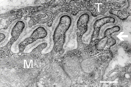
| תיאורElectron micrograph of neuromuscular junction (cross-section).jpg |
English: Electron micrograph showing a cross-section through the neuromuscular junction. T is the axon terminal, M is the muscle fiber. The arrow shows junctional folds with basal lamina. Postsynaptic densities are visible on the tips between the folds. The scale is 0.3 µm. |
| תאריך יצירה | Originally uploaded to en.wikipedia on 10 במרץ 2006. |
| מקור | Synapse Web at the National Institute of Mental Health, National Institutes of Health; originally from en.wikipedia; description page is/was here. |
| יוצר | National Institute of Mental Health; originally uploaded by Nrets at en.wikipedia. |
רישיון
| Public domainPublic domainfalsefalse |
This image is a work of the National Institutes of Health, part of the United States Department of Health and Human Services, taken or made as part of an employee's official duties. As a work of the U.S. federal government, the image is in the public domain.
 |
||
| הקובץ הזה זוהה כקובץ חופשי מכל מגבלה ידועה תחת חוק זכויות היוצרים, כולל כל הזכויות הקשורות והסמוכות. | ||
https://creativecommons.org/publicdomain/mark/1.0/PDMCreative Commons Public Domain Mark 1.0falsefalse
יומן העלאה מקורי
(All user names refer to en.wikipedia)
- 2006-03-10 20:07 Nrets 433×289×8 (97758 bytes) Electron micrograph showing a cross section through the neuromuscular junction. T is the axon terminal, M is the muscle fiber. The arrow shows junctional folds with basal lamina. Postsynaptic densities are visible on the tips between the folds. Scale is 0
כיתובים
נא להוסיף משפט שמסביר מה הקובץ מייצג
פריטים שמוצגים בקובץ הזה
מוצג
10 במרץ 2006
היסטוריית הקובץ
ניתן ללחוץ על תאריך/שעה כדי לראות את הקובץ כפי שנראה באותו זמן.
| תאריך/שעה | תמונה ממוזערת | ממדים | משתמש | הערה | |
|---|---|---|---|---|---|
| נוכחית | 06:41, 22 במרץ 2007 | | 289 × 433 (95 ק"ב) | Fran Rogers | {{Information |Description=Electron micrograph showing a cross section through the neuromuscular junction. T is the axon terminal, M is the muscle fiber. The arrow shows junctional folds with basal lamina. Postsynaptic densities are visible on the tips be |
שימוש בקובץ
הדף הבא משתמש בקובץ הזה:
שימוש גלובלי בקובץ
אתרי הוויקי השונים הבאים משתמשים בקובץ זה:
- שימוש באתר ar.wikipedia.org
- שימוש באתר cs.wikipedia.org
- שימוש באתר de.wikipedia.org
- שימוש באתר es.wikipedia.org
- שימוש באתר fa.wikipedia.org
- שימוש באתר gl.wikipedia.org
- שימוש באתר ko.wikipedia.org
- שימוש באתר pt.wikipedia.org
- שימוש באתר ru.wikipedia.org
- שימוש באתר uk.wikipedia.org
- שימוש באתר zh.wikipedia.org
מטא־נתונים
קובץ זה מכיל מידע נוסף, שכנראה הגיע ממצלמה דיגיטלית או מסורק שבהם הקובץ נוצר או עבר דיגיטציה.
אם הקובץ שונה ממצבו הראשוני, כמה מהנתונים להלן עלולים שלא לשקף באופן מלא את הקובץ הנוכחי.
| כיוון מצלמה | רגילה |
|---|
.jpg)