File:Symptoms_of_fibromyalgia.png
Uit Wikipedia, de vrije encyclopedia
Oorspronkelijk bestand (730 × 688 pixels, bestandsgrootte: 155 kB, MIME-type: image/png)
| Dit is een bestand van Wikimedia Commons. Onderstaande beschrijving komt van de beschrijving van het bestand daar. |
Inhoud
Beschrijving
| BeschrijvingSymptoms of fibromyalgia.png |
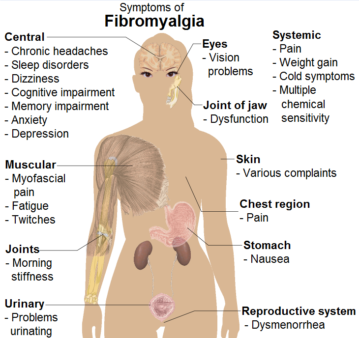
English: Common signs and symptoms of fibromyalgia. (See Wikipedia:Fibromyalgia#Signs and symptoms).
Model: Mikael Häggström. To discuss image, please see Template talk:Human body diagrams
References
|
| Datum | |
| Bron | All used images are in public domain. |
| Auteur |
When using this image in external works, it may be cited as:
or
|
| Andere versies |
 |

|
Bestand:Symptoms of fibromyalgia.svg is een vectorversie van dit bestand. Indien niet van slechtere kwaliteit dient deze gebruikt te worden in plaats van deze rasterafbeelding.
File:Symptoms of fibromyalgia.png → File:Symptoms of fibromyalgia.svg
Zie Help:SVG voor meer informatie.
|
 |
Licentie
| Public domainPublic domainfalsefalse |
| Ik, de auteursrechthebbende van dit werk, geef dit werk vrij in het publieke domein. Dit is wereldwijd van toepassing. In sommige landen is dit wettelijk niet mogelijk; in die gevallen geldt: Ik sta iedereen toe dit werk voor eender welk doel te gebruiken, zonder enige voorwaarden, tenzij zulke voorwaarden door de wet worden voorgeschreven. |
Human body diagramsMain article at: Human body diagrams Template location:Template:Human body diagrams How to derive an imageDerive directly from raster image with organsThe raster (.png format) images below have most commonly used organs already included, and text and lines can be added in almost any graphics editor. This is the easiest method, but does not leave any room for customizing what organs are shown. Adding text and lines: Derive "from scratch"By this method, body diagrams can be derived by pasting organs into one of the "plain" body images shown below. This method requires a graphics editor that can handle transparent images, in order to avoid white squares around the organs when pasting onto the body image. Pictures of organs are found on the project's main page. These were originally adapted to fit the male shadow/silhouette.
Organs:
Derive by vector templateThe Vector templates below can be used to derive images with, for example, Inkscape. This is the method with the greatest potential. See Human body diagrams/Inkscape tutorial for a basic description in how to do this.
Examples of derived works
Licensing
|


.png)


Bijschriften
Items getoond in dit bestand
beeldt af
19 apr 2009
image/png
Bestandsgeschiedenis
Klik op een datum/tijd om het bestand te zien zoals het destijds was.
| Datum/tijd | Miniatuur | Afmetingen | Gebruiker | Opmerking | |
|---|---|---|---|---|---|
| huidige versie | 12 dec 2009 08:50 | | 730 × 688 (155 kB) | Mikael Häggström | +jaw joint |
| 11 dec 2009 17:52 |  | 730 × 688 (152 kB) | Mikael Häggström | Replaced myself with the female shadow, since it affects more females than males, with a ratio of approximately 9:1 | |
| 19 apr 2009 09:48 |  | 777 × 675 (430 kB) | Mikael Häggström | spelling | |
| 19 apr 2009 09:38 |  | 777 × 675 (430 kB) | Mikael Häggström | {{Information |Description={{en|1=g}} |Source=Own work by uploader |Author=Mikael Häggström |Date=g |Permission= |other_versions= }} <!--{{ImageUpload|full}}--> |
Bestandsgebruik
Geen enkele pagina gebruikt dit bestand.
Globaal bestandsgebruik
De volgende andere wiki's gebruiken dit bestand:
- Gebruikt op en.wikipedia.org
- Gebruikt op en.wikiversity.org
- Gebruikt op is.wikibooks.org
- Gebruikt op it.wikipedia.org
- Gebruikt op simple.wikipedia.org
Metadata
Dit bestand bevat metadata met EXIF-informatie, die door een fotocamera, scanner of fotobewerkingsprogramma toegevoegd kan zijn.
| Horizontale resolutie | 37,79 dpc |
|---|---|
| Verticale resolutie | 37,79 dpc |