Datei:Red_White_Blood_cells.jpg
From Wikipedia, the free encyclopedia
Kei höcheri Uflösig verfüegbar.
Red_White_Blood_cells.jpg (500 × 326 Pixel, Dateigrößi: 57 KB, MIME-Typ: image/jpeg)
| Die Datei un d Informatione derzue were us em zäntrale Mediearchiv Wikimedia Commons (Allmänd) iibunde. | Zur Bschriibigssite uff de Allmänd |
Bschryybig
| BschryybigRed White Blood cells.jpg |
العربية: صورة بالميكروسكوب الالكتروني لخلية دم حمراء(يسار), صفيحة دموية(وسط)و خلية دم بيضاء(يمين)
Azərbaycanca: Soldan sağa: eritrosit, trombosit, leykosit
Čeština: Červená krvinka (vlevo), krevní destička (uprostřed) a bílá krvinka (vpravo)
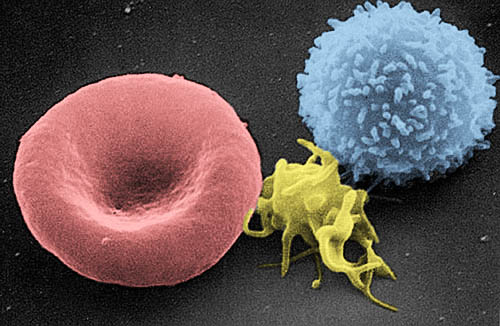
Deutsch: Rasterelektronenmikroskop (REM)-Aufnahme, eingefärbt: Erythrozyt, Thrombozyt, Leukozyt (von links nach rechts)
English: Colored Scanning electron microscope (SEM)-picture: erythrocyte, thrombocyte, leukocyte (from left to right:)
Dansk: Fra venstre mod højre: erythrocyt, thrombocyt, leukocyt
Magyar: Színezett Pásztázó elektronmikroszkóp kép: eritrocita, trombocita, leukocita (balról jobbra)
Italiano: Da sinistra verso destra: eritrocita, piastrina e linfocita T
日本語: 左から: 赤血球、血小板、白血球。
Español: De izquierda a derecha: eritrocito, trombocito, linfocito T
Suomi: Väritetty pyyhkäisyelektronimikroskooppikuva, jossa vasemmalta oikealle: punasolu, verihiutale ja valkosolu.
Français : De gauche à droite : érythrocyte, thrombocyte et leucocyte
Nederlands: rode en witte bloedcel, en in het midden een bloedplaatje
Polski: Od lewej do prawej: erytrocyt, trombocyt, leukocyt
Română: De la stânga la dreapta: globulă roşie de sânge, trombocită, limfocită
Русский: Слева направо: эритроцит, тромбоцит, лейкоцит
Português: Da esquerda para a direita : eritrócito, plaqueta e leucócito
中文:由左至右:紅血球,血小板,白血球
A three-dimensional ultrastructural image analysis of a T-lymphocyte (right), a platelet (center) and a red blood cell (left), using a Hitachi S-570 scanning electron microscope (SEM) equipped with a GW Backscatter Detector. |
|||||||
| Datum | 21. Septämber 2004 (Original-Hochladedatum) | |||||||
| Quälle | [1] | |||||||
| Urheber | Electron Microscopy Facility at The National Cancer Institute at Frederick (NCI-Frederick) | |||||||
| Gnähmigung (Wyternutzig vu däre Datei) |
|
|||||||
| Anderi Versione |
Abgleiteti Wärk vo dere Datei: |
|||||||

This image was copied from wikipedia:nl where it was uploaded by nl:User:Svdmolen on 21 sep 2004 10:38. The original description was:
rode en witte bloedcel - http://web.ncifcrf.gov/ - PD
Kurzbeschreibungen
Ergänze eine einzeilige Erklärung, was diese Datei darstellt.
Tiết
صورة بالميكروسكوب الالكتروني لخلية دم حمراء(يسار), صفيحة دموية(وسط)و خلية دم بيضاء(يمين).
In dieser Datei abgebildete Objekte
Motiv
scanning electron micrograph Änglisch
Herstellungsmethode Tüütsch
Rasterelektronenmikroskopie Tüütsch
Dateiversione
Klick uf e Zytpunkt zu aazeige, wie s dert usgsäh het.
| Version vom | Vorschaubild | Mäß | Benutzer | Kommentar | |
|---|---|---|---|---|---|
| aktuell | 18:07, 3. Jun. 2016 | | 500 × 326 (57 KB) | Jakob Suckale | Scanning EM picture was colorized to highlight the different cell types. Red blood cell in red, platelet in yellow, lymphocyte in blue. |
| 18:05, 3. Jun. 2016 |  | 500 × 326 (57 KB) | Jakob Suckale | Scanning EM picture was colorized to highlight the different cell types. Red blood cell in red, platelet in yellow, lymphocyte in blue. | |
| 22:44, 9. Nov. 2005 |  | 500 × 326 (36 KB) | E rulez | This image was copied from wikipedia:nl. The original description was: rode en witte bloedcel - http://web.ncifcrf.gov/ - PD {| border="1" ! date/time || username || edit summary |---- | 21 sep 2004 10:38 || Svdmolen || <nowiki>(rode en witte bloedcel - |
Verwändig vu dr Datei
Wältwyti Dateinutzig
Die andere Wikis bruche die Datei:
- Gebruch uf ang.wikipedia.org
- Gebruch uf arc.wikipedia.org
- Gebruch uf ar.wikipedia.org
- Gebruch uf ar.wikiversity.org
- Gebruch uf arz.wikipedia.org
- Gebruch uf ast.wikipedia.org
- Gebruch uf bar.wikipedia.org
- Gebruch uf be.wikipedia.org
- Gebruch uf bg.wikipedia.org
- Gebruch uf bn.wikipedia.org
- Gebruch uf bn.wikibooks.org
- Gebruch uf bs.wikipedia.org
- Gebruch uf ca.wikipedia.org
- Gebruch uf ckb.wikipedia.org
- Gebruch uf cs.wikipedia.org
- Gebruch uf da.wikipedia.org
- Gebruch uf de.wikipedia.org
Wältwyti Verwändig vu däre Datei aaluege.
Metadate
Die Datei het wyteri Informatione, allwäg vor Digitalkamera oder vom Scanner wo se het gschaffe.
We die Datei isch veränderet worde, de cha's sy, das die zuesätzlechi Information für di verändereti Datei nümm richtig zuetrifft.
| Breiti | 500 px |
|---|---|
| Längi | 326 px |
| Bit pro Farbkomponente |
|
| Pixelzämmesetzig | RGB |
| Orientierung | Normal |
| Aazahl vu dr Komponente | 3 |
| Horizontali Uflesig | 200 dpi |
| Vertikali Uflesig | 200 dpi |
| Software | Adobe Photoshop CS6 (Macintosh) |
| Spycherzytpunkt | 16:38, 3. Jun. 2016 |
| Exif-Version | 2.21 |
| Farbruum | Nit kalibriert |
| Eidytigi Chännig vum Orginaldokumänt | 98064A1F5B7E7BED1939FB9098746DC2 |
| Digitalisierigszytpunkt | 18:07, 3. Jun. 2016 |
| Datum, wu d Metadate s letscht Mol gänderet wore sin | 18:38, 3. Jun. 2016 |