קובץ:PET-image.jpg
ויקיפדיה האנציקלופדיה encyclopedia
גודל התצוגה המקדימה הזאת: 679 × 600 פיקסלים. רזולוציות אחרות: 272 × 240 פיקסלים | 543 × 480 פיקסלים | 869 × 768 פיקסלים | 1,132 × 1,000 פיקסלים.
לקובץ המקורי (1,132 × 1,000 פיקסלים, גודל הקובץ: 139 ק"ב, סוג MIME: image/jpeg)
| זהו קובץ שמקורו במיזם ויקישיתוף. תיאורו בדף תיאור הקובץ המקורי (בעברית) מוצג למטה. |
תקציר
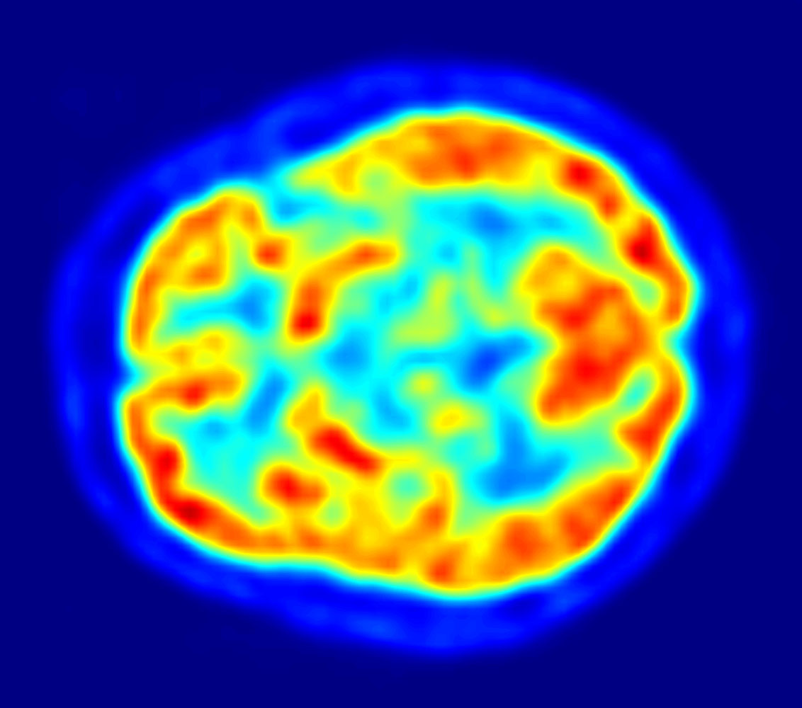
| תיאורPET-image.jpg |
English: This is a transaxial slice of the brain of a 56 year old patient (male) taken with positron emission tomography (PET). The injected dose have been 282 MBq of 18F-FDG and the image was generated from a 20 minutes measurement with an ECAT Exact HR+ PET Scanner. Red areas show more accumulated tracer substance (18F-FDG) and blue areas are regions where low to no tracer have been accumulated.
العربية: صورة مقطعية للدماغ البشري تظهر استهلاك الطاقة. |
|||
| תאריך יצירה | ||||
| מקור | נוצר על־ידי מעלה היצירה | |||
| יוצר | Jens Maus (http://jens-maus.de/) | |||
| אישורים והיתרים (שימוש חוזר בקובץ זה) |
|
כיתובים
נא להוסיף משפט שמסביר מה הקובץ מייצג
صورة مقطعية للدماغ البشري تظهر استهلاك الطاقة.
פריטים שמוצגים בקובץ הזה
מוצג
היסטוריית הקובץ
ניתן ללחוץ על תאריך/שעה כדי לראות את הקובץ כפי שנראה באותו זמן.
| תאריך/שעה | תמונה ממוזערת | ממדים | משתמש | הערה | |
|---|---|---|---|---|---|
| נוכחית | 05:00, 12 בדצמבר 2017 | | 1,000 × 1,132 (139 ק"ב) | SteinsplitterBot | Bot: Image rotated by 270° |
| 17:36, 16 במרץ 2010 |  | 1,132 × 1,002 (134 ק"ב) | Damato | uploaded another PET image with a higher resolution which might be more usable for printing it and which has a better color scale. | |
| 12:47, 7 בנובמבר 2005 |  | 405 × 373 (48 ק"ב) | Damato | This is an image taken from a typical PET acquisition. It is a tomographic view of a brain examination in transaxial view. Red areas show more accumulated radioactivity and blue areas are partions where low to no activity was accumulated. It should illust |
שימוש בקובץ
הדפים הבאים משתמשים בקובץ הזה:
שימוש גלובלי בקובץ
אתרי הוויקי השונים הבאים משתמשים בקובץ זה:
- שימוש באתר ar.wikipedia.org
- שימוש באתר arz.wikipedia.org
- שימוש באתר ast.wikipedia.org
- שימוש באתר bg.wikipedia.org
- שימוש באתר bn.wikipedia.org
- שימוש באתר ca.wikipedia.org
- שימוש באתר de.wikipedia.org
- שימוש באתר de.wikibooks.org
- שימוש באתר el.wikipedia.org
- שימוש באתר en.wikipedia.org
- Positron emission tomography
- Neurolinguistics
- Human brain
- Scintigraphy
- Timeline of tuberous sclerosis
- User:Portakalsinatra
- Wikipedia:Wikipedia Signpost/2011-03-07/Features and admins
- User talk:Silver seren/Archive 10
- Childhood acquired brain injury
- User:Rkasinadhuni3/practice sandbox
- User:Mcorrin3/Sandbox Practice
- User:LoriJeanMarie/Brain science practice page
- User:Gilyardterence/Pediatric Acquired Brain Injury
- Wikipedia:Wikipedia Signpost/Single/2011-03-07
- Wikipedia:WikiProject Cannabis/Members
- User:Anthonyhcole/Parkinson's disease
- User:Silver seren/Barnstars
- User:Flyer22 Frozen/Human brain
- User:Cglife.bmarcus/WikiProjectCards/WikiProject Cannabis
- שימוש באתר en.wikiquote.org
- שימוש באתר en.wikiversity.org
- שימוש באתר es.wikipedia.org
מטא־נתונים
קובץ זה מכיל מידע נוסף, שכנראה הגיע ממצלמה דיגיטלית או מסורק שבהם הקובץ נוצר או עבר דיגיטציה.
אם הקובץ שונה ממצבו הראשוני, כמה מהנתונים להלן עלולים שלא לשקף באופן מלא את הקובץ הנוכחי.
| כיוון מצלמה | רגילה |
|---|---|
| רזולוציה אופקית | 600 dpi |
| רזולוציה אנכית | 600 dpi |
| תוכנה בשימוש | Adobe Photoshop CS3 Macintosh |
| התאריך והשעה של שינוי הקובץ | 15:34, 16 במרץ 2010 |
| מרחב הצבע | sRGB |
| רוחב התמונה | 1,002 px |
| גובה התמונה | 1,132 px |
| התאריך והשעה של הפיכת הקובץ לדיגיטלי | 16:34, 16 במרץ 2010 |
| תאריך השינוי האחרון של המטא־נתונים | 16:34, 16 במרץ 2010 |
אוחזר מתוך "https://he.wikipedia.org/wiki/קובץ:PET-image.jpg"