Dosiero:Anatomy_of_Human_Ear_with_Cochlear_Frequency_Mapping.svg
From Wikipedia, the free encyclopedia

Grando de tiu PNG antaŭprezento de tiu SVGa dosiero: 674 × 519 rastrumeroj. Aliaj distingivoj: 312 × 240 rastrumeroj | 624 × 480 rastrumeroj | 998 × 768 rastrumeroj | 1 280 × 986 rastrumeroj | 2 560 × 1 971 rastrumeroj.
Bildo en pli alta difino (SVG-dosiero, 674 × 519 rastrumeroj, grandeco de dosiero: 33 KB)
| Jen dosiero de la Wikimedia-Komunejo. La priskribo en ties priskriba paĝo estas montrata suben.
|
Resumo
| PriskriboAnatomy of Human Ear with Cochlear Frequency Mapping.svg |
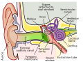
English: The human ear and frequency mapping in the cochlea. The three ossicles incus, malleus, and stapes transmit airborne vibration from the tympanic membrane to the oval window at the base of the cochlea. Because of the mechanical properties of the basilar membrane within the snail-shaped cochlea, high frequencies will produce a vibration peak near the oval window, whereas low frequencies will stimulate receptors near the apex of the cochlea (locations for three frequencies indicated schematically). Information from the cochlear receptor cells is transmitted to the cochlear nuclei via the 8th cranial nerve, and on through the midbrain to the cortex. |
| Dato | |
| Fonto | Propra verko (Originala teksto: Own work by uploader, derived from File:Anatomy_of_the_Human_Ear.svg ) |
| Aŭtoro | Inductiveload |
| Permeso (Reuzo de la dosiero) |
Ĉi tiu dosiero estas disponebla laŭ la permesilo Krea Komunaĵo Atribuite-Samkondiĉe 2.5 Ĝenerala.
|
| Ceteraj versioj |
[redakti]
|
| SVG genesis InfoField | This file is translated using SVG switch elements: all translations are stored in the same file. |


Titoloj
Donu unulinian priskribon de la enhavo de ĉi tiu dosiero
A labelled cross-sectional diagram of the human ear.
A labelled cross-sectional diagram of the human ear.
15 feb. 2009
image/svg+xml
4c58b13c29ef1eb6b3ba8c18aed2cd55519d51d1
34 114 bajto
519 rastrumero
674 rastrumero
Dosierhistorio
Alklaku iun daton kaj horon por vidi kiel la dosiero tiam aspektis.
| Dato/Horo | Bildeto | Grandecoj | Uzanto | Komento | |
|---|---|---|---|---|---|
| nun | 21:29, 16 sep. 2018 | | 674 × 519 (33 KB) | JoKalliauer | added systemLanguage="eo" |
| 17:21, 16 sep. 2018 |  | 674 × 519 (32 KB) | JoKalliauer | added systemLanguage="de" | |
| 05:33, 11 sep. 2018 |  | 674 × 519 (87 KB) | Jmarchn | Bigger (proportional real size) and full redraw (more realistic) of the auricle. Ossicles in white colour. Eardrum with contour. Added 3 labels. Add fundus to the bone and subcutaneous tissues, add superior auricular muscle, add transparency to middle ear, add separation between middle and inner ear, add division to internal auditory canal. | |
| 13:40, 29 apr. 2009 |  | 800 × 600 (98 KB) | Inductiveload | swap incus/malleus | |
| 15:10, 15 feb. 2009 |  | 800 × 600 (98 KB) | Inductiveload | {{Information |Description={{en|1=The human ear and frequency mapping in the cochlea. The three ossicles incus, malleus, and stapes transmit airborne vibration from the tympanic membrane to the oval window at the base of the cochlea. Because of the mechan |
Dosiera uzado
La jenaj paĝoj ligas al ĉi tiu dosiero:
Suma uzado de la dosiero
La jenaj aliaj vikioj utiligas ĉi tiun dosieron:
- Uzado en en.wikipedia.org
- Uzado en en.wikibooks.org
- Uzado en he.wikipedia.org
- Uzado en lt.wikipedia.org
- Uzado en meta.wikimedia.org
- Uzado en www.wikidata.org
Metadatumoj
Ĉi tiu dosiero entenas aldonan informon, probable aldonitan de la diĝita fotilo aŭ skanilo uzita por ĝin krei aŭ diĝitigi. Se la dosiero estas modifita disde sia originala stato, detaloj povas ne ĝuste priskribi tiun modifitan bildon.
| Larĝeco | 674.23 |
|---|---|
| Alteco | 518.84 |